Evolutionary Morphology of Primate Hands and Feet
Funding: NSF BCS 1317525, 1440742, 1552848
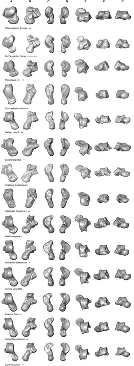
Above: Early euprimate tarsals compared to those of extant
strepsirrhines. On left, cheirogaleids;
on right, fossil adapiforms. Top row shows articulated ankles (actual size) in
dorsal view. Note extreme size difference between Marcgodinotius from Gujarat (G)
in India (~53 mya) and Notharctus
(~52 mya). Middle row elements scale to tuber+posterior astragalar facet length
(dashed lines). Note that Marcgodinotius
has the shortest distal calcaneal arm of the four, including similarly sized,
non-leaping Cheirogaleus major (Gebo,
1987)
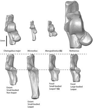
Above: Plot of fibular facet angles for various taxa. Distal views of two primate astragali show landmarks used in measurement. From Boyer et al. (2010b)
|
Intellectual Merit: The information that is contained in the morphology of primate upper ankle joints (astragalus and calcaneus bones) provides a window into taxonomic affinities, locomotor mode, and evolutionary history. The PI and co-PI propose to analyze this system to contribute evidence towards several central questions relating to primate origins and evolution. This work will have excellent potential to yield novel insight due to comprehensive taxonomic sampling, employment of new methods for detailed morphological quantification, and the use of phylogenetically-informed statistical analyses. Specifically, four major questions will be addressed: 1) What amount of adaptation to hindlimb-propelled leaping occurred during the transition from stem primates to crown primates? 2) At what point in various basal euprimate clades did significant locomotor transitions occur? 3) To what degree, and in what ways did ancestral adapiforms, omomyoids, anthropoids, strepsirrhines and tarsiers differ from each other in overall and specific features of the upper ankle joint? and 4) What does new and recently described material of basal anthropoids and “protoanthropoids” from the Late Eocene of the Fayum depression, Asia, and India tell us about early anthropoid evolution and anthropoid affinities to other Euprimates? The development of methods for comprehensively and repeatably quantifying this richly informative anatomical system has remained a major challenge to students in the field. Furthermore, the utility of qualitative comparisons using published imagery is often limited simply by the difficulty for achieving consistent orientation and adequate feature resolution for these complexly variable, and often miniscule, elements. As more fossil primate ankles are described and the diversity of morphologies increases, the task for a researcher of traveling to view all relevant specimens becomes untenable, and the reliance on published imagery and measurements (rather than access to actual specimens) becomes greater. The necessity of effective morphological quantification also is magnified because maintaining personal knowledge of the increasing number of relevant forms is becoming unmanageable. The PI and co-PI propose to mitigate these issues by producing and digitally publishing a 3-D dataset of primate astragali and calcanei. An extant comparative dataset and many fossils are already scanned. They will employ this digital imagery to evaluate hypotheses relating to the functional and phylogenetic significance of morphological variation, and (in an explicit phylogenetic context) to reconstruct the morphotypes of basal euprimates and various euprimate clades using 1) a set of 58 detailed measurements they have previously published, 2) additional new measurements they are developing, and 3) geometric morphometric datasets. In the project description, the PI’s use a pilot sample to concretely demonstrate that these techniques have already brought resolution to certain questions left open by traditional techniques. Broader Impacts: This project provides funding for a graduate student, and provides training for a diverse group of students at Brooklyn College, which is primarily an undergraduate institution. All students will receive mentoring in scientific research and its communication to the public. Students will be trained in use of 3-D computer visualization techniques. Because such techniques are also applied in various industries and represent critical resume items even for undergraduates who do not pursue a career in academia and move into fields as diverse as medicine, engineering, art or architecture. Because this work is so focused on the early history of primates, it will lead to a better understanding of what it means for humans to have an evolutionary relationship with other animals, especially for students with a limited background in evolution and paleoanthropology. A major goal of this project is the dissemination of 3D data it generates to other researchers and educators through online databanks that incorporate educational tools like Morphobrowser. |
Phylogenetic utility of tarsal bones
|
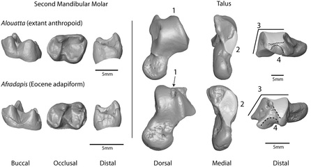
Seiffert et al. (2009) analogized Afradapis, an Eocene adapiform that exhibits “anthropoid-like” dental and gnathic features, with Alouatta because of teeth reflecting folivory. Note that there are essentially no distinctive molar features contra-indicating platyrrhine status for the fossil. On the other hand, the newly discovered astragalus of Afradapis (Boyer et al. 2010b) can be easily recognized as characteristically “strepsirrhine,” while that of Alouatta is distinctively “anthropoid,” even though both bones also clearly indicate that these taxa shared a fairly similar locomotor habit (e.g., a shallow body in medial view). Surprisingly, such ankle features typically receive little attention in phylogenetic analyses (e.g., Ni et al., 2007; Seiffert et al., 2009), where dental characters outnumber ankle characters at rates of 10:1 or more. Labeled features: 1) Superior astragalar foramen never retained in known anthropoids; 2) Medial tibial facet reduced in anthropoids; 3) Fibular facet vertical in anthropoids; 4) Flexor fibularis groove (lower dashed line) plantar to proximal margin of lateral tibial facet (upper dashed line) in haplorhines, instead of lateral to it as in strepsirrhines.
|
|